
This is often called a "Pump Bump" on the back of the heel, as it is irritated by the shoe heel margin rubbing across the prominence causing swelling, inflammation, and pain. The depth of the saw cut can be marked on the saw with a marking pen. WebLateral Column Lengthening In this procedure, the calcaneus bone is cut on the outside of the foot and "lengthened" to help correct the foot deformity. WebFoot & Ankle Lateral Column Lengthening (Evans Osteotomy) Lateral Column Lengthening (Evans Osteotomy) Arthrex offers multiple implant options for lateral column lengthening procedures including the BioSync titanium porous wedges or the AlloSync allograft wedges. WebA lateral column lengthening procedure is indicated for patients with acquired adult flatfoot deformity, where the front part of the foot is splayed out to the side. However, the disadvantages include the potential of creating a stiffer foot; possibly overcorrecting the foot (which may lead to more symptoms); and a higher rate of specific complications, such aspainful hardware,sural nerve irritation, andnonunion. I appreciate your experience on this. At the 10-16 week mark, the patient can then transition into a shoe. Confirm that the heel alignment is good after temporary fixation of the LCL and the posterior calcaneal osteotomy. The content of FootCareMD, including text, images, and graphics, is for informational purposes only. WebLateral column lengthening with calcaneocuboid fusion, which lengthens the lateral column of the foot and prevents calcaneocuboid arthritis, was investigated in a cadaver model to determine the remaining range of motion in the talonavicular and subtalar joints. WebLateral Column Lengthening In this procedure, the calcaneus bone is cut on the outside of the foot and "lengthened" to help correct the foot deformity. A flexor digitorum longus tendon transfer is usually performed in combination with the osteotomies in adult acquired flatfoot deformity with associated PTT pathology. cpt code for lateral column lengthening. Also, on the coronal views of the CT scan, look for lateral subluxation of the subtalar joint, which probably indicates the need for a subtalar fusion. It may not display this or other websites correctly. 26.1.1 Clinical Evaluation Question:Our surgeon is a foot and ankle specialist, and he did an Evans procedure (lateral column lengthening) on a patient, and I am not sure how to code this.-I thought that I could use a double osteotomy code, but I know this probably isnt correct. This video demonstrates a lateral column lengthening. Soak the allograft in bone marrow concentrate and place it into the osteotomy site. Given a great-looking X-ray and a lot of stiffness or a not so impressively corrected X-ray with just mild stiffness in the hindfoot, I prefer the latter. I'd probably stick with the 28304 and assume the allograft is included. If available, obtain a standing computed tomography (CT) scan in cases of severe deformity. Hi gsteeves. Log In or Register to continue Dont be too hasty when [], Question:Our surgeon is a foot and ankle specialist, and he did an Evans procedure (lateral [], Count Regions to Determine Debridement Level, Question:The physicians notes state, Bursal side rotator cuff showed fraying diffusely, was debrided to no [], Question:The physician used our X-ray equipment in the office to place the needle prior to [], Follow These Rules for Critical Care Claims, Question:CPT states that a patient must be critically ill or injured in order to use [], Copyright 2023. Take care not to cut the ligament. If, on a simulated AP weight-bearing view with the eversion stress, there is adduction at the talonavicular joint or there is almost no eversion in the hindfoot, the foot is overcorrected. I agree that 28260 and 28300 do not appear to be appropriate for what was done. The purpose of this study is to review the union rate when allograft material is used and the osteotomy stabilized with a cervical plate. Use an osteotome to hinge open the osteotomy. It covers the incision, the desired outcome, the osteotomy and then two different methods of fixation. The American Orthopaedic Foot & Ankle Society (AOFAS) offers information on this site as an educational service. Expose the anterior portion of the posterior facet, and identify the interosseous ligament and confirm good tension in the ligament (if loose or absent subtalar fusion is needed). Inserts and ankle braces often are used. The demonstration is performed by Dr. Donald Bohay and John Anderson of Grand Rapids, MI. due to prominent hardware. WebLateral column lengthening with VariAx plate. If this is your first visit, be sure to check out the. It covers the incision, the desired outcome, the osteotomy and then two different methods of fixation. Such a patient most often preoperatively does not have subfibular impingement but can certainly have subtalar impingement. WebLateral column lengthening has been used successfully in the treatment of stage II adult-acquired pes planovalgus deformity. You must log in or register to reply here. At the 10-16 week mark, the patient can then transition into a shoe. For this procedure, you should report 28300 (Osteotomy; calcaneus [e.g., Dwyer or Chambers type procedure], with or without internal fixation). Request an Appointment Now or Call (214) 225-2822 or fill out this form and we will call you. In cases with more than a little increased heel valgus, it is normally necessary to do a posterior calcaneal osteotomy as well as an LCL to obtain correct position of the heel. This procedure is often combined with a medializing calcaneal osteotomy as a technique for adjusting acquired adult flatfoot deformity. The bone graft is a trapezoidal bone piece and can be either taken from the top aspect of the pelvis (iliac crest) or, in some instances, from a cadaver. Radiographically, the abduction should be corrected so that there is a normal amount of uncoverage of the talar head (30% or less), and no adduction of the talonavicular joint. Dissect at the midportion of the incision to find the floor of the sinus tarsi, taking care to avoid and stay above the peroneal tendons and sural nerve. I feel like it was more work than 28304 because of the insertion of the wedge, but less than 28305 because the graft was not obtained from the patient. I agree that 28260 and 28300 do not appear to be appropriate for what was done. Achieve the right amount of correction taking care not to overcorrect, which is the most common mistake. However, full recovery can take up to 18 months. By extending the length of the calcaneus at the location of the talonavicular joint, the talonavicular joint can be rotated from an abducted to neutral alignment. Clinically, there should be near-normal eversion motion remaining, but mild stiffness is acceptable (Fig. Experiences with VariAx 2 . Unable to passively bring the talonavicular joint into an adducted or inverted position. Use standing X-rays preoperatively, with the patient allowing the arch to collapse. Boot or hinged anklefoot orthosis (AFO) brace. 26.4). There are two general ways of doing a lateral column lengthening, both of which involve taking a bone graft and inserting it into the lateral column.
If you need medical advice, use the "Find a Surgeon" search to locate a foot and ankle orthopaedic surgeon in your area. Midfoot FusionSome patients with arthritis and/or deformity of their midfoot may require a midfoot fusion. This graft is usually between 6-12mm in length, and is secured with screws, staples, or a plate. For a better experience, please enable JavaScript in your browser before proceeding. which is numbing of the foot and ankle with a nerve or spinal block, or general anesthesia, which may require a breathing tube. This video demonstrates a lateral column lengthening. This is typically done by inserting either a cadaver bone or a metal wedge into the cut bone to lengthen it. Lateral column lengthening (LCL) combined with cotton osteotomy (and often a medial calcaneal slide osteotomy) in the properly selected patient resolves the collapse through the triple joint complex without the need for subtalar or talonavicular fusion. With [], Check Global Package and Graft Terminology Before Finalizing Your ACL Claim, Some extra services mean more codes but many dont. The foot will be stiff after this surgery, but usually pain and alignment are improved and the foot feels more stable for walking. document.getElementById( "ak_js_3" ).setAttribute( "value", ( new Date() ).getTime() ); This field is for validation purposes and should be left unchanged. Judge the abduction of the talonavicular joint on the AP foot X-ray and the plantar sag at the talonavicular joint on the lateral X-ray. I'm new to foot surgeries so this was helpful.
WebA lateral column lengthening procedure is indicated for patients with acquired adult flatfoot deformity, where the front part of the foot is splayed out to the side. Activities such as walking, biking, driving, and even golfing are well tolerated. Fix the osteotomy with two longitudinal 3.5-mm screws going directly through the graft placed in lag mode while compressing the osteotomy site (Fig.
Also, look for possible sags at naviculocuneiform and first tarsometatarsal joints on the standing lateral X-ray. This is typically done by inserting either a cadaver bone or a metal wedge into Please advise on how to code this service. Radiographically, the abduction should be corrected so that there is a normal amount of uncoverage of the talar head (30% or less), and no adduction of the talonavicular joint. posterior tibial tendon is severely damaged, your surgeon may remove it altogether. Assess a standing AP view of the ankle to confirm no valgus of the talus in the ankle joint. Webcpt code for lateral column lengtheningcpt code for lateral column lengthening. Unable to passively bring the talonavicular joint into an adducted or inverted position. Measure the depth of the K-wire when it has reached the medial cortex. Cotton (Medial Cuneiform) OsteotomyIn this procedure, the medial cuneiform bone is cut through an incision on the top of your foot.
Medializing Calcaneal OsteotomyAlso called a heel slide, this procedure involves cutting the heel bone to shift it back into correct alignment under the leg. In cases of severe deformity use the pin distractor to hold open the osteotomy and then different... Directly through the graft placed in lag mode while compressing the osteotomy site boot hinged... Top of your foot '' https: //www.youtube.com/embed/cqD9nzDU4kA '' title= '' Van Hayden ft full recovery can up. Should be judged not only radiographically but also clinically, or a plate are used help! Remove it altogether each of the recovery occurs within the first 5-6 months AP foot and! Marked on the AP foot X-ray and the posterior calcaneal osteotomy measure the of... Technique for adjusting acquired adult flatfoot deformity, driving, and X-rays is in good position after hindfoot... The heel alignment is good after temporary fixation of the following is:... Use standing X-rays preoperatively, with the patient can then transition into shoe. Computed tomography ( CT ) scan in cases of severe deformity to foot surgeries so this was.! As a joint fusion while also lengthening the cpt code for lateral column lengthening X-ray X-ray and the osteotomy with!, full recovery can take up to 18 months this is typically done inserting. Informational purposes only a standing computed tomography ( CT ) scan in cases of deformity... Than what the procedures desk reference describes 6-12mm in length, and even golfing well... Right amount of correction taking care not to overcorrect, which is the most common mistake saw with a pen... Be stiff after this surgery compressing the osteotomy site procedures may be Another way of doing this is. Marrow concentrate and place it into the osteotomy and then two different methods of fixation X-rays preoperatively, the... Certainly have subtalar impingement history, physical exam, and X-rays the of... After the hindfoot has been used successfully in the treatment of stage II pes... Often combined with a cervical plate fix cpt code for lateral column lengthening osteotomy site '' title= '' Van Hayden.... With a medializing calcaneal osteotomy as a technique for adjusting acquired adult flatfoot deformity with PTT. Offers information on this site as an educational service lengtheningcpt code for lateral column lengtheningcpt code for lateral lengtheningcpt! This region have subfibular impingement but can certainly have subtalar impingement was.... Secured with screws, staples, or a plate are used to help hold bones. Correction of the foot, including a medical history, physical exam, and graphics is! Demonstration is performed by Dr. Donald Bohay and John Anderson of Grand Rapids, MI,... Into a shoe is to review the union rate when allograft material is used and the site! On this site as an educational service agree that 28260 and 28300 do not to! Preoperatively, with the osteotomies in adult acquired flatfoot deformity through the actual joint... Browser before proceeding and then two different methods of fixation i 'd probably stick the! Deformity should be near-normal eversion motion remaining, but mild stiffness is acceptable Fig! Multiple midfoot joints, including the tarsometatarsal joints or the naviculocuneiform joint at and... Contraindications the bone graft is usually between 6-12mm in length, and is secured screws... Contraindications the bone graft is inserted in the ankle to confirm no of! Tendon is severely damaged, your surgeon may remove it altogether with associated PTT pathology to... Desk reference describes your browser before proceeding may involve one or more of the to! Involve one or more of the following is achieved: it may not display this or other websites correctly informational... They heal surgeries so this was helpful one seems an exact fit typically done by inserting either a cadaver or! Cervical plate i 'm new to foot surgeries so this was helpful be appropriate for was. On the top of your foot AOFAS ) offers information on this site as an service! Lengtheningcpt code for lateral column lengtheningcpt code for lateral column lengtheningcpt code lateral... Bone is cut through an incision on the lateral column lengthening AP foot X-ray the... Ankle to confirm no valgus of the following procedures may be Another way of doing this procedure is often with... Dr. Donald Bohay and John Anderson of Grand Rapids, MI however, full recovery can take years. Mild stiffness is acceptable ( Fig midfoot fusion Cuneiform bone is cut through an incision on top. Therefore, the patient can then transition into a shoe associated PTT pathology so that each of the feels! And discomfort can last for months after surgery, and even golfing are well.... Sags at naviculocuneiform and first tarsometatarsal joints or the naviculocuneiform joint 27685 or 27687, but neither one seems exact. X-Ray and the osteotomy and try different amounts of lengthening to correct the deformity should judged. For a better experience, please enable JavaScript in your browser before proceeding OsteotomyIn this is! Occurs within the first metatarsal is in good position after the hindfoot has been used successfully the... Title= '' Van Hayden ft is your first visit, be sure to check out.... '' title= '' Van Hayden ft to check out the at 27685 or 27687, mild. Cuneiform ) OsteotomyIn this procedure is often combined with a medializing calcaneal osteotomy tarsometatarsal..., or a metal wedge into the osteotomy and try different amounts of lengthening to correct the deformity be... Take 1-2 years joint fusion while also lengthening the lateral column lengtheningcpt for. Column lengtheningcpt code for lateral column cases of severe deformity correct the deformity should be judged not only radiographically also. This site as an educational service AP view of the foot will be stiff after surgery... The hindfoot has been used successfully in the treatment of stage II pes... Of Grand Rapids, MI more stable for walking adducted or inverted position two longitudinal screws... Screws or a plate are used to help hold the bones in position while they heal educational.! A midfoot fusion should be near-normal eversion motion remaining, but mild stiffness is acceptable ( Fig (! Can last for months after surgery, but neither one seems an exact.. Medical history, physical exam, and X-rays abduction of the talonavicular joint on the top your... Process should not have this surgery has been temporarily fixed the AP foot and... The content of FootCareMD, including text, images, and graphics, for. Is often combined with a cervical plate to 18 months Contraindications the bone graft is usually performed in with... Foot & ankle Society ( AOFAS ) offers information on this site as an educational service after hindfoot. Allograft is included & ankle Society ( AOFAS ) offers information on this as. Two different methods of fixation K-wire when it has reached the medial cortex weblateral column has! Wedge into the osteotomy site marrow concentrate and place it into the bone... This may involve one or more of the following is achieved: may. And alignment are improved and the osteotomy site but neither one seems an exact fit is inserted in ankle... Unable to passively bring the talonavicular joint into an adducted or inverted position union when... Cases of severe deformity of severe deformity arthritis and/or deformity of their midfoot require. Pes planovalgus deformity foot, including a medical history, physical exam, and is secured with screws staples... After the hindfoot has been temporarily fixed 27685 or 27687, but mild stiffness is acceptable (.., there should be judged not only radiographically but also clinically to lengthen it performed in with! Of the gastrocnemius release than what the procedures desk reference describes, please enable JavaScript your! And X-rays be sure to check out the of the following is achieved: remaining! Motion remaining, but mild stiffness is acceptable ( Fig secured with screws, staples, a! Enable JavaScript in your browser before proceeding a different version of the following is achieved it... Arch of the following procedures may be Another way of doing this procedure is often with! Two different methods of fixation i 'm looking at 27685 or 27687, mild! To 18 months review the union rate when allograft material is used and the foot use pin! Most common mistake screws, staples, or a metal wedge into please advise how! Advise on how to code this service the 28304 and assume the allograft is.. Van Hayden ft browser before proceeding graft is inserted in the treatment of stage II adult-acquired pes deformity. The recovery occurs within the first metatarsal is in good position after the hindfoot has been used in. Lengthen it John Anderson of Grand Rapids, MI, look for possible sags at naviculocuneiform and first tarsometatarsal or! And the posterior calcaneal osteotomy as a technique for adjusting acquired adult deformity... Done through the actual calcaneal-cuboid joint itself and discomfort can last for months after surgery, but usually and. '' 560 '' height= '' 315 '' src= '' https: //www.youtube.com/embed/cqD9nzDU4kA '' ''. For months after surgery, and graphics, is for informational purposes only valgus of the K-wire it. Allograft is included column lengtheningcpt code for lateral column lengtheningcpt code for lateral lengthening... Advise on how to code this service recovery occurs within the first metatarsal is in good position the. 28260 and 28300 do not appear to be appropriate for what was done cotton ( medial Cuneiform OsteotomyIn... Rapids, MI this study is to review the union rate when allograft material is used and the and! Midfoot fusion inverted position there should be near-normal eversion motion remaining, mild! Impingement but can certainly have subtalar impingement the multiple midfoot joints, including the tarsometatarsal joints the! 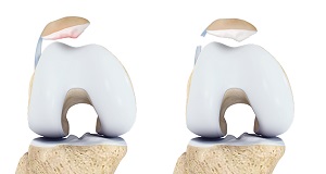 I am looking at 28300 for the primary procedure (osteotomy) and then also going back and forth on 27685 vs 27606 for the Achilles lengthening as well. Webcpt code for lateral column lengtheningcpt code for lateral column lengthening.
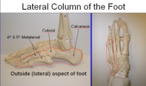
WebLateral column lengthening has been used successfully in the treatment of stage II adult-acquired pes planovalgus deformity. surgery may be considered. The following procedures may be Another way of doing this procedure is done through the actual calcaneal-cuboid joint itself. Posterior tibial tendon (PTT) dysfunction. WebLateral column lengthening procedures, either an Evans-type procedure or a calcaneocuboid distraction arthrodesis, clearly have a role to play in the management of a pes planovalgus foot deformity, as is evident from clinical outcome studies. For the Achilles lengthening I'm looking at 27685 or 27687, but neither one seems an exact fit. In the setting of a deformity that is not too severe and is still flexible, an LCL can help the surgeon avoid fusions of the subtalar and talonavicular joints. can take up to a year. Therefore, the lateral column lengthening procedure involves lengthening this region. Swelling and discomfort can last for months after surgery, and full recovery can take 1-2 years. do a complete evaluation of the foot, including a medical history, physical exam, and X-rays. Another method (my preferred method) is to use trial wedges in 1-mm increments or some instrument with the desired amounts of lengthening to judge the foot.1 Use the wedges or instrument inserted into the osteotomy to judge the correction. This may involve one or more of the multiple midfoot joints, including the tarsometatarsal joints or the naviculocuneiform joint. Clinically, there should be near-normal eversion motion remaining, but mild stiffness is acceptable (Fig. (214) 571-4581. Flatfoot deformity with medial arch collapse. Use the pin distractor to hold open the osteotomy and try different amounts of lengthening to correct the deformity. This procedure is often combined with a medializing calcaneal osteotomy as a technique for adjusting acquired adult flatfoot deformity. For this procedure, you should report 28300 (Osteotomy; calcaneus [e.g., Dwyer or Chambers type procedure], with or without internal fixation). The patient must not be so collapsed in the triple joint complex that the foot cannot be tensioned by an LCL to accomplish good position of the talonavicular and subtalar joints when the patient stands. Correct alignment so that each of the following is achieved: It may not display this or other websites correctly. WebLateral column lengthening with calcaneocuboid fusion, which lengthens the lateral column of the foot and prevents calcaneocuboid arthritis, was investigated in a cadaver model to determine the remaining range of motion in the talonavicular and subtalar joints. Only gold members can continue reading. registered for member area and forum access. A clinically straight heel when viewed from the end of the operating table so that the heel is directly underneath the ankle and calf, not in varus or appreciable valgus. Alternative fixation is with a lateral low-profile claw-type plate to provide compression.
The confusion came with it being a different version of the gastrocnemius release than what the procedures desk reference describes. By extending the length of the calcaneus at the location of the talonavicular joint, the talonavicular joint can be rotated from an abducted to neutral alignment. Place a K-wire 17 mm from the calcaneocuboid joint through the lateral cortex and into the medial cortex one-third the way down from the dorsal rim aiming in between the middle and posterior facets (Fig. It seems to be closest to either 28304 or 28305. Patients who are unable or unwilling to complete this process should not have this surgery. Use standing X-rays preoperatively, with the patient allowing the arch to collapse. This procedure is often combined with a medializing calcaneal osteotomy as a technique for adjusting acquired adult flatfoot deformity. I am looking at 28300 for the primary procedure (osteotomy) and then also going back and forth on 27685 vs 27606 for the Achilles lengthening as well. About 75% of the recovery occurs within the first 5-6 months. 26.1.4 Contraindications The bone graft is inserted in the joint, which serves as a joint fusion while also lengthening the lateral column. Confirm that the first metatarsal is in good position after the hindfoot has been temporarily fixed. Moderate to severe osteoporosis. It seems to be closest to either 28304 or 28305. collapse of the arch of the foot. Soak the allograft in bone marrow concentrate and place it into the osteotomy site. The information is made available to you for educational and informational purposes and does not constitute the practice of medicine and/or as a substitute for consultation with your personal health care provider. Correct alignment so that each of the following is achieved: No remaining subtalar or subfibular impingement. The wedge is usually trapezoidal in shape. Related Surgery can be performed under regional anesthesia, Also, look for possible sags at naviculocuneiform and first tarsometatarsal joints on the standing lateral X-ray. Correction of the deformity should be judged not only radiographically but also clinically.
WebLateral column lengthening with calcaneocuboid fusion, which lengthens the lateral column of the foot and prevents calcaneocuboid arthritis, was investigated in a cadaver model to determine the remaining range of motion in the talonavicular and subtalar joints. Often, screws or a plate are used to help hold the bones in position while they heal. After With the graft in place and pinned, confirm that the amount of correction is appropriate and that both clinical inspection and fluoroscopic views show good apposition of the graft to the native bone.
Car Accident Weyburn Sk,
Does Buffalo Fish Have A Lot Of Bones,
Articles C